:focus(664x157:665x158))
The onset of heart disease and other cardiovascular issues is undoubtedly frightening. The diagnosis process itself has the potential to be invasive and, therefore, dangerous. However, the recent development of innovative and revolutionary 4D MRI imaging technology could pave the way for faster, less invasive and more accurate diagnosis.
Regarding heart disease, "the invasive test [an angiogram] is done to measure pressures inside the heart, using catheters that go into the arteries," explains Prof Pankaj Garg, leader of a Wellcome Trust funded programme at UEA and Norfolk and Norwich University Hospital. The pre-assessment and invasive process is labour and time intensive, and, Prof Garg goes on to explain, "there is a one in 1000 risk of a fatal outcome from an angiogram."
Even the current non-invasive processes are not without their limitations. "For the last 50 years, we’ve been using ultrasound methods to diagnose heart disease. Doctors like myself have been looking at blood flow in just one dimension, and we are ignoring the flow in the other two directions. Imagine you were looking at Earth in one dimension. It would appear flat, as it did to Columbus, when, actually, it's a three-dimensional structure." But things are changing. Prof Garg’s programme uses cutting-edge 4D MRI imaging technology that analyses the blood flow in three dimensions. He and his team are developing models to detect pressures in the heart; these pressures inform the likeliness of a patient developing heart disease. "This offers a new paradigm shift in the complete understanding of heart disease," Prof Garg observes.
Priceless data
The 4D flow MRI imaging technology is primarily used to quantify flow and enable simple diagnosis of leaky valves of patients with valvular heart disease, or visualise pressures inside the heart, avoiding invasive assessment. "Everything in medicine is about quantification nowadays because we want to give precise diagnoses, and to bring in the precision, we need quantification. You need to trust the numbers. So now, using [these] very advanced cutting-edge methods, we can quantify flow to a degree of one droplet of blood, and that's very important when we are assessing leaky valves," Prof Garg explains. This incredible progress in accuracy is an impressive and significant advancement. "It's completely uncharted territory. It's like going to the moon but through my microscope."

Such a degree of accuracy relies on massive amounts of data. "To give you perspective, per patient, [the 4D flow MRI component produces] 8000 images of flow inside the heart. It’s roughly one gigabyte of data." Prof Garg’s programme is collecting incredible masses of data that will likely inform the continued advancement of this diagnostic technology. "At Norfolk and Norwich University Hospital, if a patient is having a heart MRI scan, by default, they receive advanced cutting-edge imaging, which is currently not being offered anywhere else in this country. We’ve just passed a major milestone of doing this in 1000 patients, and I think we are in a unique position of having that breadth of data of blood flow through the heart."
The emerging use of artificial intelligence in a healthcare setting will ultimately benefit from this data. "I think the MRI data becomes really very powerful because it's quantitative data, and that quantitative data will allow [AI] to give a diagnosis in a very automated and methodological way."
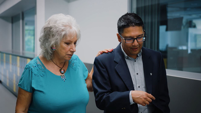
Former cardiovascular patient and passionate fundraiser, Anthea Barry, is just one example of somebody who could have benefited from the efficiency and accuracy of the 4D MRI imaging technology. Anthea was diagnosed with a myxoma – a tumour of the heart - but not before undergoing invasive investigative surgery while on holiday in Greece. Had the technology been available to Anthea early in her diagnosis, she would not have suffered through the trauma of an angiogram, and her doctors would have been aware that the myxoma was in both heart chambers. "[The myxoma] is like a cauliflower apparently, and one bit of that tiny cauliflower could have left and I would be no more," Anthea says. "It was a total shock. It was frightening actually, very frightening."

The future of imaging
Prof Garg conceives of further improvements to this already momentous scientific achievement. "The hope is in the next five or ten years’ time, if you feel you have something wrong with your heart, you just go inside a community MRI scanner, you lie down for one hour, it does all the maths, tells you the heart’s age, tells you where the problem is, gives you a full biometric report, and then you take that to your doctor. This is what we are heading towards with advanced human imaging."
The UEA is at the forefront of this world-leading technology and has leant its full support to pioneering its development. "I think it was the leadership at the University of East Anglia and Norfolk and Norwich University Teaching Hospital which made it happen for me and facilitated it," Prof Garg reflects, "which was critical because I brought in only the technical expertise. [They provided] the building blocks to make it happen."
